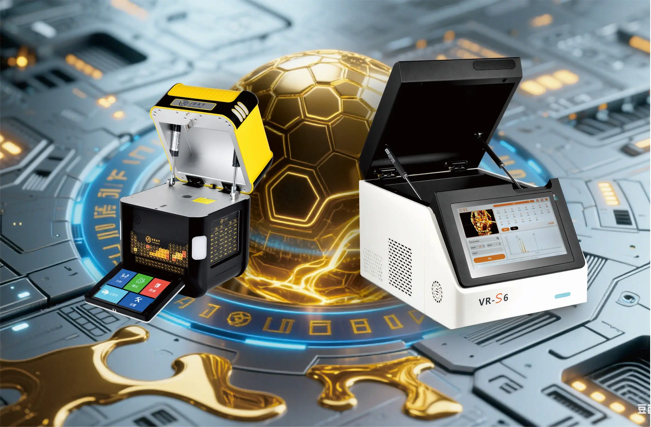
 VRAY仪器有限公司
You rely on the x-ray tube in a xrf spectrometer as the main x-ray source that excites your samples during x-ray fluorescence analysis. This system lets you investigate the elemental composition of materials using a non-destructive analytical technique. When you use a x-ray fluorescence spectrometer, the x-ray tube generates primary x-rays that trigger fluorescence signals from your sample. These signals help you achieve accurate, non-destructive elemental analysis. Advances in x-ray tube technology, such as those in the VRAY XRF ANALYZER, have boosted performance and lifespan, making the system more reliable and cost-effective. For example, the global market for x-ray tubes in XRF spectrometers reached $1.2 billion in 2023 and is expected to grow further:
Market Metric | 价值 / Description |
|---|---|
Market Size (2023) | USD 1.2 billion |
Market Projection | |
CAGR (2024-2033) | 5.8% |
You benefit from this technology because it enables rapid, precise, and safe elemental analysis in many industries.
Key Takeaways
这 x-ray tube is the main source of x-rays in an XRF spectrometer, exciting samples to reveal their elemental makeup without damage.
You can adjust the tube’s voltage, current, and target material to match different samples and improve analysis accuracy.
Key parts of the tube include the cathode (electron source), anode (x-ray target), and housing (vacuum and safety shield).
Proper tube settings and beam focusing help get clear, precise results while protecting users from radiation.
Following safety rules and regular maintenance keeps the x-ray tube working well and ensures safe operation.
X-ray Tube in a XRF Spectrometer
Source of Primary X-rays
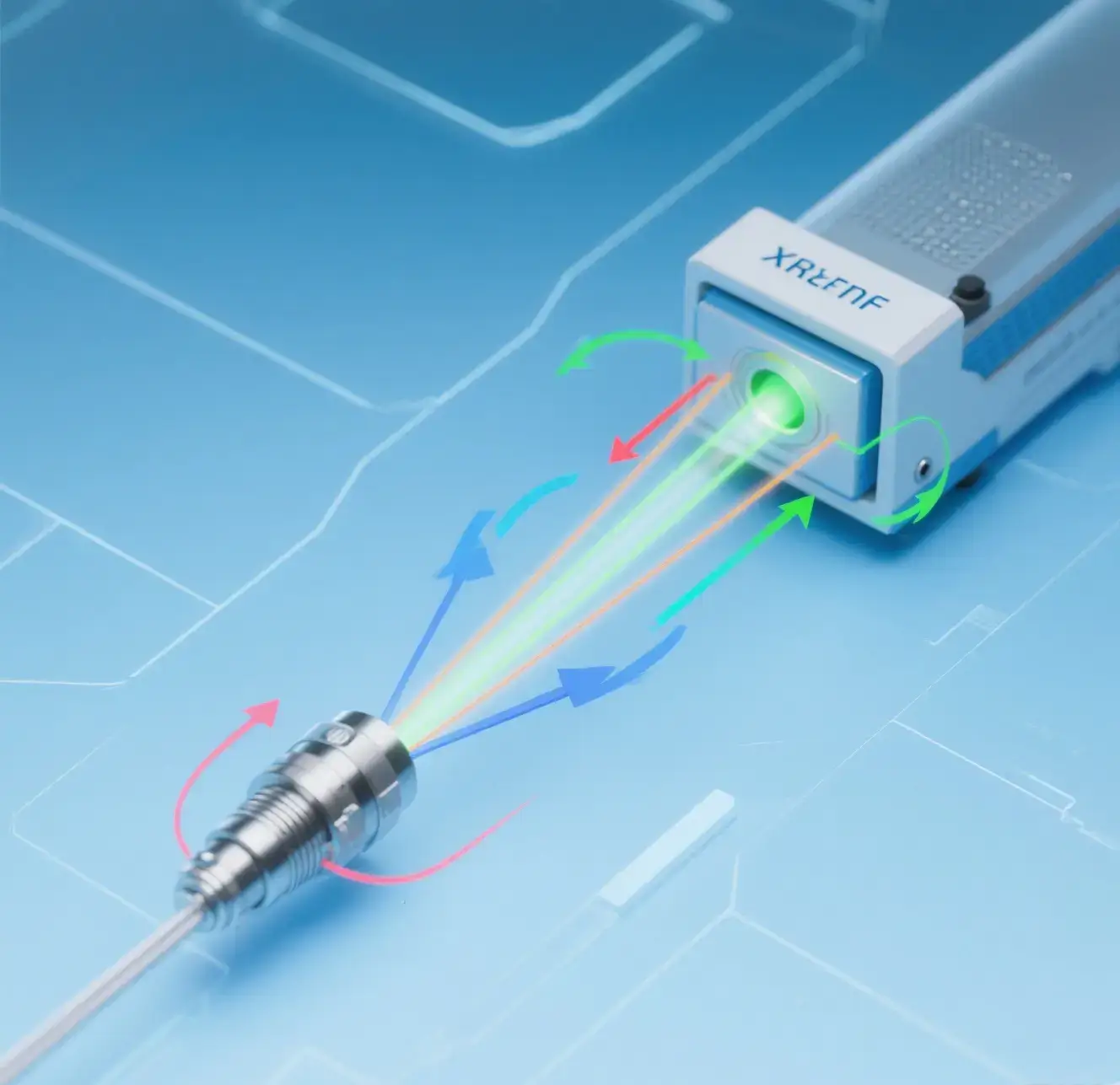
You depend on the x-ray tube in a XRF光谱仪 as the heart of your analysis system. This device acts as the primary x-ray source, producing the high-energy beam needed to excite your sample. When you power up the spectrometer, the x-ray generator inside the tube creates a focused stream of electrons. These electrons hit the anode, which then emits x-rays. The beam travels through the tube window and reaches your sample, starting the process of x-ray fluorescence.
The x-ray tube in a xrf spectrometer gives you control over the intensity and energy of the x-ray beam. You can adjust the voltage and current to match the needs of your sample. This flexibility lets you analyze a wide range of materials, from metals to plastics. The x-ray generator also keeps the process safe by containing the high-voltage parts and shielding you from stray radiation.
You can see how different tube features affect your results in the table below:
Parameter | Description | Relevance to XRF Effectiveness |
|---|---|---|
Target Materials | 钨, molybdenum, chromium, 银, rhodium; each produces x-rays of different energies. | Enables selection of appropriate x-ray energy for element analysis. |
Power Range | 10 自 4000 watts; higher power yields stronger x-rays with greater penetration. | Allows tuning of x-ray intensity for different sample types. |
Voltage Range | 30 kV to 100 kV; higher voltage produces higher energy x-rays. | Facilitates analysis of materials with varying depth and density. |
Mechanical Structure | Side-window, end-window, transmission target types. | Different emission geometries suit various XRF applications. |
Focal Spot Size | Small for high resolution; large for high intensity. | Balances resolution and intensity for optimal XRF performance. |
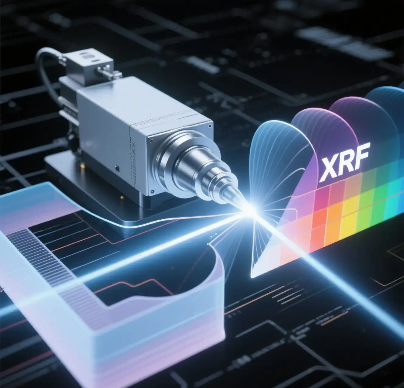
Excitation in X-ray Fluorescence
When you use a x-ray fluorescence spectrometer, the x-ray tube sends a beam of primary x-rays onto your sample. This beam excites the atoms in the material. The energy from the x-ray generator knocks electrons out of their inner shells. As the atoms return to their normal state, they emit characteristic x-rays. These fluorescent x-rays carry information about the elemental composition of your sample.
You rely on this process for accurate surface analysis and deep material studies. The x-ray tube in a xrf spectrometer lets you choose the right beam energy and intensity for your application. For example, you can select a tungsten or molybdenum target to match the elements you want to detect. The system uses a collimated beam to focus the x-rays, which improves the precision of your measurements.
You benefit from innovations in x-ray tube design. New anode materials like gold allow higher tube voltages, which help you detect heavy elements such as cadmium. Graphene detector windows make it possible to measure lighter elements like fluorine. Electronic control systems balance peak shaping and dead time, giving you clearer spectra. Onboard computing power now supports advanced spectrum deconvolution and element quantification, making your results faster and more reliable.
The x-ray tube in a xrf spectrometer works together with the detector and electronics to give you a complete picture of your sample. You can use this technology in many fields, such as mining, environmental testing, 和质量控制. 这 energy dispersive x-ray fluorescence analyzer and other xrf spectrometers rely on the x-ray generator to produce the characteristic x-ray fluorescence signals you need for precise analysis.
X-ray Tube Components
Cathode and Filament
You find the cathode and filament at the core of every x-ray tube. The filament, usually made of tungsten, heats up when you pass an electric current through it. This heating causes the filament to emit electrons. The cathode acts as the source for these electrons. When you apply a high voltage, the electrons accelerate toward the anode. The number of electrons released depends on the filament current. More current means more electrons and a stronger x-ray output. You control the filament current to adjust the intensity of the x-ray beam for your analysis.
Anode (目标)
The anode, also called the target, sits opposite the cathode inside the x-ray tube. When the fast-moving electrons hit the anode, they slow down quickly. This sudden stop produces x-rays. The material you choose for the anode, such as molybdenum or tungsten, affects the energy and type of x-rays created. Heavier elements in the anode give you stronger and more specific x-ray lines. You can select the target material to match the elements you want to analyze in your sample. The anode must also handle a lot of heat, so cooling systems often keep it from overheating during use.
Tube Housing
这 tube housing surrounds the cathode and anode, creating a safe and controlled environment. You find that the housing keeps air out by maintaining a vacuum. This vacuum lets electrons travel freely from the cathode to the anode. The housing also includes a thin beryllium window. This window allows the generated x-rays to exit the tube and reach your sample without much loss of energy. The housing shields you from stray radiation and helps direct the x-ray beam exactly where you need it. Some housings use special shapes or materials to focus the beam for better results.
这 cathode, anode, and housing work together to generate and direct x-rays efficiently. You control the settings and choose the right materials to get the best performance from your x-ray tube.
X-ray Generation in XRF
Electron Acceleration
You start the process by powering up the x-ray generator. Inside the tube, the filament heats up and releases electrons. A strong electric field accelerates these electrons toward the anode. When the electrons hit the target, they lose energy quickly. This energy loss creates x-ray photons. The way you set up the target area and its distance from the crystal changes how many x-rays you get. For example, experiments show that smaller target areas and closer distances increase x-ray intensity. You can control the energy of the electrons by adjusting the target position. This helps you get a stable and efficient x-ray generator for your analysis.
XRF uses the photoelectric effect to create electron vacancies, which leads to the emission of fluorescent x-rays.
You rely on calibration standards and statistical models to reduce errors and improve accuracy.
Advances in micro-XRF and synchrotron sources give you more precise and reliable results.
X-ray Production Process
When the accelerated electrons strike the anode, you get two types of x-rays: characteristic and Bremsstrahlung. The characteristic x-rays have specific energies that match the elements in the target. Bremsstrahlung x-rays form a continuous spectrum. Your x-ray generator produces both, but you often select the target material to get the best energy for your sample. The detector picks up the fluorescent x-rays that your sample emits after being hit by the primary beam. Research shows that changing the geometry of the target and the electron path can boost the efficiency of x-ray production. Computer simulations help you understand how electrons move and focus inside the tube.
Collimation and Beam Focus
You need to focus the x-ray beam to get accurate results. Collimators shape the beam and limit its size. A smaller aperture gives you better spatial resolution but takes more time to scan. If you use a large aperture, the detector may pick up signals from outside your target, which lowers accuracy. A focused pencil-beam collimator can improve localization accuracy to less than 0.6 mm. The table below shows how different collimator settings affect your analysis:
Parameter/Aspect | Description/Effect |
|---|---|
Collimator aperture size | Smaller apertures improve spatial resolution (~1 mm) but increase scan time. |
Detector collimator aperture | Large apertures cause detection of XRF signals beyond target boundaries, reducing target accuracy. |
Beam focus (pencil-beam collimator) | 2 mm FWHM excitation beam improves localization accuracy (<0.6 mm offset) and enhances CNR and DSC. |
Spatial resolution (FWHM) | ~7.0 mm with large detector collimator aperture. |
Localization accuracy | Improved to <0.6 mm offset with focused pencil-beam collimator. |
You can see that careful control of the beam and detector settings helps you get the most accurate and sensitive results from your x-ray fluorescence analysis.
Tube Settings and XRF Results
Target Material Choice
You can select different target materials in your x-ray tube to match your analysis needs. Each material, such as tungsten, molybdenum, or silver, produces x-rays with unique energies. This choice helps you focus on certain elements in your sample. For example, using a copper filter can boost the signal for chromium detection and lower the background noise. A study found that a copper filter between 100 μm and 140 μm increased the signal-to-noise ratio for chromium, making it easier to detect even small amounts. This adjustment lowered the detection limit to 0.32 mg/L, which meets strict environmental standards. By choosing the right target and filter, you improve both sensitivity and accuracy in xrf.
Voltage and Current Effects
You control the voltage and current settings on your x-ray tube to change the energy and intensity of the x-ray beam. Higher voltage increases the energy of the x-rays, which helps you analyze heavier elements or look deeper into samples. Higher current boosts the number of x-rays, making your measurements faster and more reliable. 然而, you must balance these settings. If you use too much power, the tube can overheat and wear out faster. Manufacturer data shows that running at high current with low voltage can shorten filament life by up to three times. The temperature of the tube rises quickly with more power, which can cause cracks and reduce performance over time.
Voltage and current changes also affect the quality of your results:
Higher voltage can increase background noise for light elements.
Higher current usually improves the signal but may not always help with accuracy.
Impact on Measurement Accuracy
Your tube settings have a direct impact on measurement accuracy in xrf. Small changes in voltage or current can shift the signal-to-noise ratio (SNR) for different elements. The table below shows how SNR changes for several elements when you adjust tube voltage and current:
Parameter Variation | Element | Effect on Median SNR (因素) | Notes |
|---|---|---|---|
Tube voltage increase 30 kV → 50 kV | Si | 0.57 | SNR decreased due to increased background and decreased peak area for lighter elements. |
Tube voltage increase 30 kV → 50 kV | K | 0.85 | Slight decrease in SNR. |
Tube voltage increase 30 kV → 50 kV | Ti | 0.98 | Nearly unchanged SNR. |
Tube voltage increase 30 kV → 50 kV | Rb | 3.05 | Significant increase in SNR. |
Tube voltage increase 30 kV → 50 kV | Zr | 1.85 | Substantial increase in SNR. |
Tube current increase 30 mA → 50 马 | Si | 1.26 | Moderate increase in SNR. |
Tube current increase 30 mA → 50 马 | K | 1.21 | Moderate increase in SNR. |
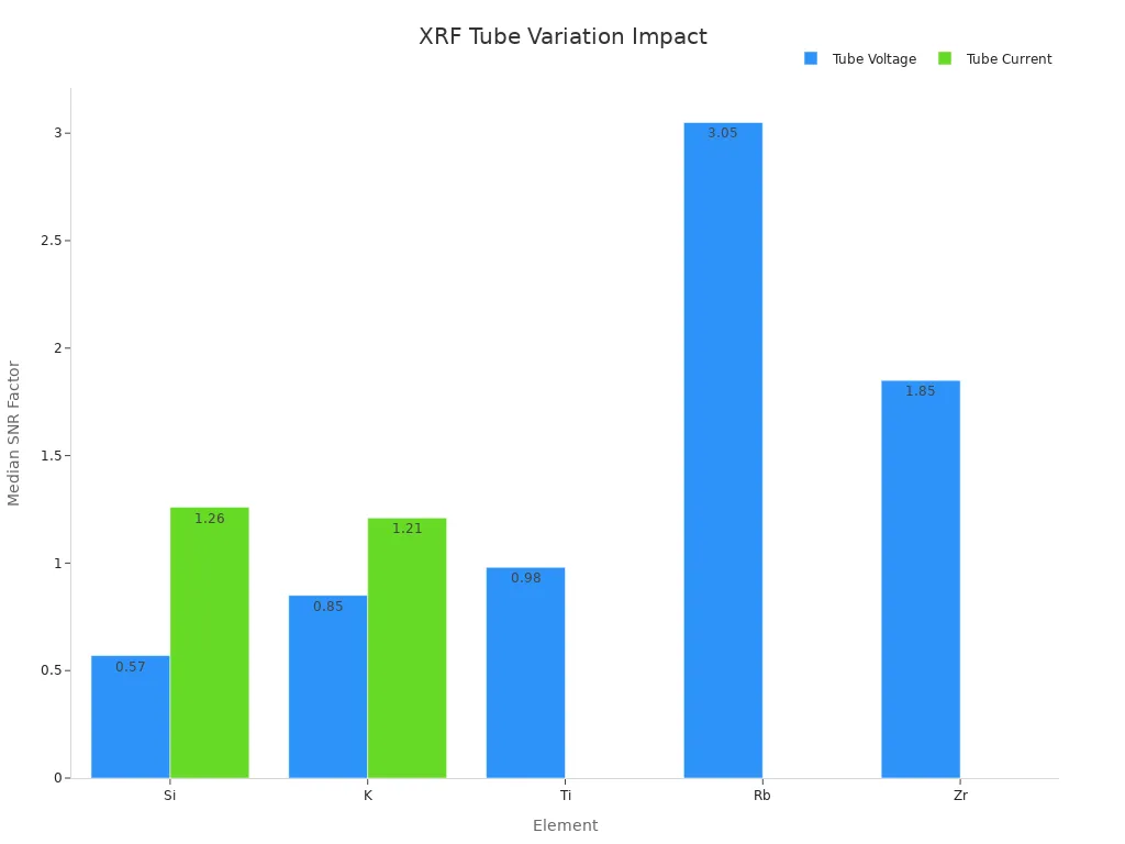
You see that increasing voltage helps with heavier elements like rubidium and zirconium but can hurt the SNR for lighter elements like silicon. Current increases give a moderate boost for most elements. For best results, you should match your tube settings to your sample and the elements you want to measure. This approach improves both qualitative and quantitative analysis, especially when you need accurate surface analysis or a clear spectrum.
Note: Studies show that using element ratios, such as Fe/Ca, can reduce errors caused by tube setting differences and sample changes. This method helps you compare results between different labs and instruments.
Safety and Maintenance
Safe Operation
You must always follow strict safety rules when working with an x-ray tube in an XRF光谱仪. The primary x-ray beam can deliver a high dose rate, sometimes over 4,000 rads per hour. Even though the cabinet design keeps radiation leakage below 0.5 milliroentgens per hour at 5 cm from any surface, you should never place your hands or eyes near the beam source. Always keep a safe distance from the nose of the device and use benchtop stands for low-density samples to reduce backscatter exposure.
Never bypass safety interlocks or attempt repairs on your own.
Make sure warning lights and audible signals work before you start the spectrometer.
Only operate the device when all doors and panels are closed and locked.
Keep at least 3 feet between the spectrometer and other people.
Point the device away from people and set up a safe zone around the equipment.
Regulations require that you check for warning labels near controls and ports. These labels remind you about x-ray hazards. The FDA also requires that manufacturers provide manuals with safety procedures and maintenance schedules. You should always read these manuals before using the spectrometer.
Tip: ALARA (As Low As Reasonably Achievable) means you should minimize your time near the device, maximize your distance, and never tamper with shielding.
Maintenance Tips
Regular maintenance helps your x-ray tube and detector work safely and last longer. You should inspect the indicator lights and safety interlocks every day. If you notice any problems, report them right away. Periodically check the shielding and housing for damage. Never use the spectrometer if you see cracks or loose parts.
Clean the exterior of the spectrometer and detector with a soft, dry cloth.
Schedule professional inspections after any major repairs or if you move the device.
Make sure the detector and x-ray detectors are free from dust and debris, as this can affect performance.
Keep the area around the spectrometer clear to avoid accidental bumps or spills.
Use only approved replacement parts for the detector and x-ray detectors.
Radiation Safety Offices often conduct leakage surveys when you install the device or after repairs. Continuous monitoring with area dosimeters helps ensure safe conditions. You do not need lead aprons or personal dosimeters if you use a properly shielded cabinet XRF spectrometer. Always follow the maintenance schedule in your manual to keep your detector and x-ray detectors working at their best.
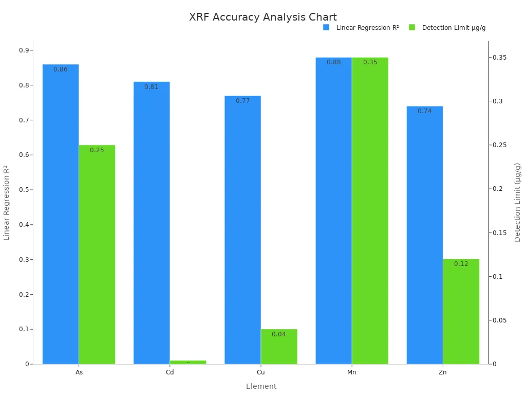
The x-ray tube stands at the core of your xrf spectrometer, enabling precise elemental analysis. You achieve high accuracy and sensitivity by selecting the right tube settings and components. Key performance factors include:
Tube power and anode material directly affect detection limits and spectral quality.
Proper operation ensures reliable results for elements from potassium to zinc.
Mo-TXRF Impact | W-TXRF Impact | |
|---|---|---|
检测限 | Lower for most elements | Better for high-Z elements |
Analytical Accuracy | High, especially for trace elements | Reliable for heavy elements |
You should always focus on tube selection, safe operation, and regular maintenance to get the best results from your xrf system.
FAQ
What happens if the x-ray tube fails during analysis?
If the x-ray tube fails, your XRF光谱仪 cannot produce x-rays. You will not get any results. You should stop using the device and contact a technician for repair or replacement.
How often should you replace the x-ray tube?
You usually replace the x-ray tube every 3 自 5 年. The exact time depends on how much you use the spectrometer and the tube settings. Always follow the manufacturer’s guidelines.
Can you clean the x-ray tube yourself?
You should not try to clean the x-ray tube. Only trained professionals should open or service the tube. Cleaning it yourself can damage the tube or expose you to radiation.
What safety gear do you need when using an XRF spectrometer?
You do not need special gear if the spectrometer has proper shielding.
Always keep your hands and face away from the x-ray window.
Never bypass safety features.
Tip: Always follow your lab’s safety rules and read the user manual.


WhatsAPP
扫描WhatsApp二维码和我们联系吧。