 أداة Vray
The discovery of x-rays by röntgen in 1895 changed science and medicine. Röntgen’s experiments gave people a way to look inside the body without surgery. His work got a lot of attention from scientists and the public. X-rays soon became very important for finding and treating diseases like cancer. The story of x-rays shows how röntgen’s idea led to fast changes in medicine. اليوم, röntgen’s work still affects technology and health.
الوجبات الرئيسية
Wilhelm Röntgen found x-rays in 1895. This let people look inside the body without surgery for the first time. Early x-rays helped doctors spot broken bones and bullets fast. They also found other problems quickly. This changed medicine forever. X-rays can be harmful if people do not follow safety rules. لذا, experts made rules to keep patients and workers safe. اليوم, x-ray technology uses digital pictures and AI. This makes scans quicker, clearer, and safer for all. X-rays are not just for medicine. They are used in security, science, and industry. This shows how much x-rays affect our daily lives.
Discovery of X-Rays
Röntgen’s Experiments
Wilhelm Conrad Röntgen found x-rays في 1895. He was working with cathode ray tubes at the time. He wanted to see how electron beams moved in low-pressure gases. Röntgen used a Crookes tube, which is a special glass tube. He covered the tube with thick black cardboard to block light. He put a screen with barium platinocyanide near the tube. When he turned on the tube, the screen started to glow. The screen glowed even though it was not in direct light. Röntgen saw that something invisible made the screen shine.
Röntgen learned that this new radiation could go through many things. It could even pass through thick books and wood. He saw that the rays made shadows of solid objects. These shadows showed shapes that normal light could not show.
Röntgen called these rays “الأشعة السينية” because he did not know what they were. He worked slowly and repeated his tests many times. His careful work helped him learn about x-rays. Other scientists had seen similar things but did not study them more.
Key steps in Röntgen’s experiments:
He used a cathode ray tube to make electron beams.
He put a fluorescent screen outside the tube.
He saw the screen glow, even when it was covered.
He tried different materials and saw x-rays could pass through most.
He named the new rays “الأشعة السينية” because he did not know what they were.
The First X-Ray Image
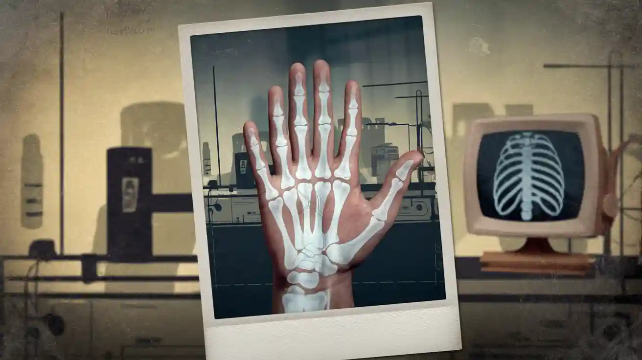
Röntgen wanted to show what x-rays could do. He decided to take a picture with the new rays. He asked his wife, Anna Bertha Röntgen, to put her hand on a photographic plate. He exposed her hand to x-rays for about 15 دقائق. The picture became the first x-ray image. The photo showed the bones in her hand and her wedding ring as a dark shadow.
This picture amazed scientists and the public. For the first time, people could see inside the body without surgery. The first x-ray image, called “Hand mit Ringen,” was a big moment in science and medicine. Doctors quickly saw that x-rays could help find broken bones or objects inside the body.
ميزة | وصف |
|---|---|
موضوع | Anna Bertha Röntgen’s left hand |
Exposure Time | 15 دقائق |
Visible Details | Bones and wedding ring |
Historical Significance | First radiograph, changed medicine |
Early Recognition
The news about x-rays spread fast. Röntgen wrote a report called “On a New Kind of Rays.” He sent it to the Würzburg Physico-Medical Society in December 1895. He also shared photos of the first x-ray image with other scientists. Newspapers in Germany and the UK soon told people about it. The public became very interested in seeing inside the body. Some people were excited, but others worried about privacy.
Scientists in many countries repeated Röntgen’s tests. They found that x-rays could help doctors find bullets or broken bones. By early 1896, hospitals in Europe and the United States started using x-rays to help doctors. Röntgen did not patent his discovery. This let others use and improve the technology for free.
The Nobel Prize committee gave Röntgen the first Nobel Prize in Physics in 1901. They praised him for finding the amazing rays that now have his name. Röntgen’s choice to share his discovery helped x-rays become important in science and medicine.
Röntgen’s discovery of x-rays changed how doctors and scientists studied the body.
The first x-ray image led to fast use and new research.
The Nobel Prize showed how important this discovery was for everyone.
Early X-Ray Adoption
Medical Uses
Doctors saw that x-rays could help patients. Hospitals started using x-ray machines soon after Röntgen’s discovery. في 1896, Massachusetts General Hospital set up an early x-ray room. Walter Dodd and Joseph Godsoe built the first x-ray machine there. By March, they made x-ray images. Early x-rays helped doctors find gunshot wounds and broken bones. They also found kidney stones and things people swallowed. These tools changed how doctors found and treated problems.
Pennsylvania Hospital got an x-ray machine in 1897. في البداية, only a few patients got x-rays. لاحقاً, more patients had x-rays done. The table below shows how x-ray use grew at Pennsylvania Hospital:
Year | All Patients with X-rays | Patients with Fractures with X-rays |
|---|---|---|
1900 | Rare use (~1.3%) | ~8.7% |
1909 | أقل من 10% (~7%) | 50% |
1920 | ~17% | 100% |
1925 | ~25% | ~86% |
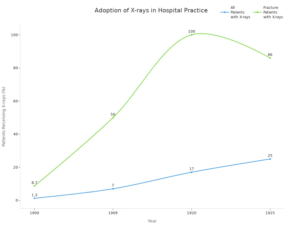
Doctors used x-rays for broken bones first. بواسطة 1920, almost every patient with a fracture got an x-ray. Hospitals made radiology departments and hired new experts. These changes helped doctors find problems and treat patients better.
Thomas Edison helped x-rays spread. He made the first fluoroscope called the Vitascope. It let people see inside the body in real time. Edison’s work made x-rays easier to use in medicine. But his helper Clarence Dally got hurt by radiation. This made people more careful about safety in radiology.
Public Fascination
People became very interested in x-rays after they were found. Newspapers wrote about the new way to see the unseen. Photographers opened studios for “bone portraits.” People could see pictures of their own bones. Public shows and talks had live x-ray demos. Volunteers watched their hands or objects appear on screens.
The fluoroscope let people see moving images inside the body.
X-rays became popular in comics and theater shows.
People thought x-rays were amazing and fun. لاحقاً, some worried about health risks. ما زال, x-rays changed society in a big way. Early use of x-rays in medicine and daily life showed how fast new ideas can change the world.
X-Ray Risks and Safety
Initial Dangers
في البداية, people did not know x-rays could be dangerous. Many doctors and scientists got hurt because they did not use protection. Some early health problems were:
Skin burns and sores showed up after using x-rays a lot.
Hair fell out where x-rays touched the skin.
Some people got cancer many years later.
Medical records showed when these injuries happened.
Photos showed burns and hair loss as proof.
Mistakes by workers and broken machines caused accidents.
Doctors and scientists learned about these dangers by looking at injuries and using tests. Court cases and expert reports showed when safety rules were not used. These early problems made people see that x-rays could be very harmful if not used safely.
“The first people who used x-rays found out the hard way that invisible rays could hurt them. What happened to them helped make things safer for everyone.”
Safety Protocols
When people saw the dangers, experts made safety rules to protect everyone from x-rays. There are three main rules for safety:
Do not spend too much time near x-rays.
Stay as far away as you can from the x-ray machine.
Wear shields like lead aprons, gloves, and glasses.
Hospitals and labs now have safety officers who check machines and teach workers. They use badges to measure how much x-ray a person gets. Regular checks and fixing machines keep them safe. The ALARA rule means people should get as little x-ray as possible.
اليوم, هناك strict rules for how much x-ray people can get. Groups like the International Commission on Radiation Protection and the FDA make sure these rules keep people safe. X-ray machines for medicine and work must meet high standards. These steps have made x-rays much safer for everyone.
Many people worry about x-rays, but studies show that if you follow the rules, the risk is very low. Learning about safety helps stop myths and keeps people safe.
X-Ray Advancements
Imaging Technology
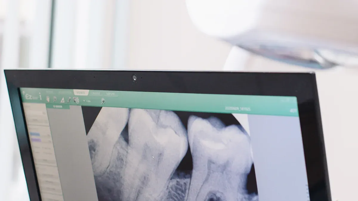
X-rays have changed a lot over time. Early x-ray machines used film to make pictures. الآن, most hospitals use digital systems instead. Digital x-ray imaging lets doctors see pictures right away on a computer. This makes things faster and easier for patients. Digital images also use less radiation. This keeps patients safer, especially kids and people who need many scans.
Modern radiology uses special detectors for clearer pictures. These detectors help doctors find problems more easily. Hospitals use portable x-ray machines for patients who cannot move. These machines work in emergency rooms and ambulances.
Digital x-rays can lower radiation by up to 80-90%. This helps keep patients and healthcare workers safe.
Doctors can now use 3D and 4D imaging to see inside the body in new ways. These tools show moving pictures and help doctors plan surgeries. Artificial intelligence (AI) helps doctors look at images quickly. AI can find problems that are hard to see. It also helps doctors work faster and make fewer mistakes.
Hospitals keep digital images in special computer systems. These systems let doctors share images with each other. This helps teams work together and helps patients get better care. New safety rules and better machines have made x-rays safer for everyone.
CT and MRI
Some of the biggest changes in radiology came with CT and MRI. CT uses x-rays and computers to make detailed pictures inside the body. ال first patient CT scan was in 1971. This tool let doctors see organs, bones, and blood vessels in 3D. CT scans got faster and clearer over time. اليوم, doctors use CT to find problems quickly and to look for diseases like cancer.
MRI does not use x-rays. It uses strong magnets and radio waves. Paul Lauterbur and Peter Mansfield helped make MRI in the 1970s. MRI gives very clear pictures of soft tissues, like the brain and heart. It is safe because it does not use radiation. MRI helps doctors find problems in the brain, spine, and joints. It is also important for cancer checks and looking at injuries.
Imaging Method | Uses X-rays? | الأفضل ل | Key Benefit |
|---|---|---|---|
CT | نعم | Bones, organs, vessels | صوم, 3D images |
MRI | لا | Brain, heart, soft tissue | No radiation, clear detail |
Both CT and MRI have changed how doctors use these tools. These imaging techniques help doctors make better choices and help patients get better.
Modern Uses
اليوم, x-rays are used in many ways in medicine and other fields. Doctors use x-rays to help with treatments, يحب biopsies and small surgeries. In emergency rooms, x-rays help find broken bones and injuries fast. CT and MRI help doctors plan cancer treatments and check if they are working.
Modern uses also include AI to read images and help with diagnosis. AI can find small changes that people might miss. Hospitals use digital systems to send images to doctors far away. This helps patients get good care, even if they live far from big hospitals.
Many hospitals now use advanced imaging to guide treatments, like draining abscesses or treating tumors without open surgery.
Outside of medicine, x-rays help in airport security and art restoration. They also check bridges and buildings for safety. Scientists use x-rays to study fossils and old objects. These new uses show how x-rays help people in many ways.
Radiology has some problems, يحب not having enough trained workers in some places. AI and new digital tools may help by making work faster and easier. Quality control and safety rules are still important to protect everyone.
X-rays are now one of the most important tools in the world. Their many uses in medicine, science, and industry show how much technology has grown since Röntgen’s discovery.
History of X-Rays: Legacy
Medical Impact
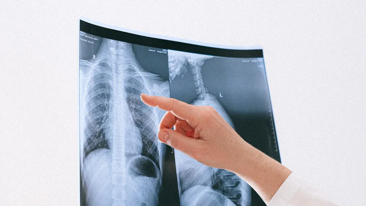
X-rays have changed health care for a long time. Doctors use x-rays every day to help patients. These images let doctors see inside the body. This helps them make better choices for care. Chest x-rays and other tests help find problems early. Finding problems early helps patients get better faster. Studies show x-rays help people live longer. Doctors can spot diseases sooner and treat them quickly.
التكنولوجيا الجديدة, like digital x-rays and AI, helps doctors a lot. AI tools help doctors find problems faster and with fewer errors. This means patients get the right care sooner. Getting care sooner can save lives. Hospitals use x-rays for many things, like broken bones and cancer treatment. These new tools show how important x-rays are for health care.
X-rays are now a key part of medical imaging. They help people everywhere get better care. New machines and digital tools bring x-rays to more places, like small clinics and urgent care centers.
Beyond Medicine
X-rays are used in many places outside hospitals. Workers use x-rays to check metal for cracks. They also use them to look at welds and make sure things are safe. Factories use x-rays to look inside circuit boards and food packages. This helps keep products safe for everyone. Scientists use x-ray crystallography to study molecules. This helped them learn about DNA’s shape.
Security teams use x-rays at airports to scan bags. They look for hidden things in luggage. Space scientists use x-ray telescopes to study stars and black holes. Archaeologists use x-rays to look at old objects without breaking them. Environmental experts use x-rays to test soil and water for pollution.
The story of x-rays has many big moments in science and technology. Labs like SLAC National Accelerator Laboratory use special x-ray tools to study materials and energy. These uses show that x-rays still help research and daily life today.
Röntgen found X-rays in 1895. This changed medicine in a big way. Scientists later made X-rays safer to use. They also invented new tools like CT and MRI. Doctors still use X-rays to find diseases today. الآن, AI helps doctors read X-ray images better. Experts think X-rays will get even clearer soon. They hope radiation will be lower and more people can get X-rays.
X-rays keep helping health care and science. Each new discovery makes their story even bigger.


واتساب
امسح رمز الاستجابة السريعة ضوئيا لبدء دردشة WhatsApp معنا.